
Threading a Brother sewing machine is a fundamental skill that every sewer must learn. While it may seem complex at first, once you understand the basic steps, you will be able to thread your machine confidently. In this guide, we …

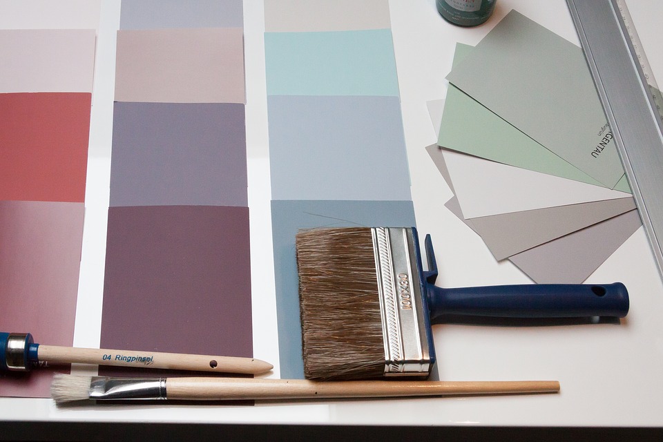
How to Choose Paint Colours That Boost Your Mood
The painters adelaide colours you choose for your home can have a significant impact on your mood and overall well-being. Whether you’re looking to create a calming retreat, energise your living space, or make your home feel more inviting, the …

Circumcision Care for a Baby
A baby who has undergone circumcision Sydney is required to be properly cared for. Use mild, unscented soap with water twice daily to clean the incision area. Avoid touching the incisions after a baby eats and poos. A baby should …

Security Guards
It is the job security services in Melbourne of a security officer to protect private property, people, or confidential information.
They also have different legal duties to different groups. First, as an individual guard, you are
legally responsible to your …

Landscape Designing: The Benefits
Landscape design is key to improving the appearance of your yard and home. Landscape
design should take into account the needs of all current retaining wall contractors Adelaide, whether they are natural or
animals. These residents need to be …
Single Parents in Painting Business
Although the Painters in Brisbane painting industry is very lucrative, you need to understand that it can also be highly
competitive, especially for single parents. Painting jobs are often seasonal and single-income
earners cannot afford to be picky. While you …
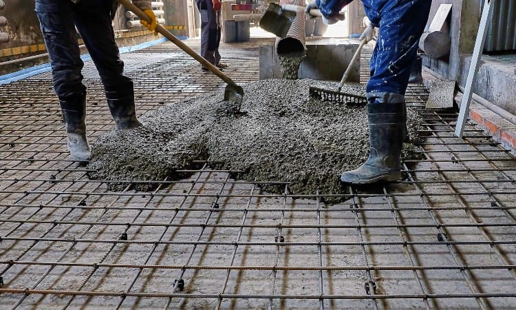
Concrete Problems and Their Solutions
Like other building materials, concrete may experience problems over time. These issues could include discolorations, shrinkage and scaling; however, they’re typically simple to manage and resolve.
Cracked Concrete
Though concrete is generally durable material, it still can become damaged over …

Types of Martial Arts
There are many types of martial arts. We will be talking about Kendo (Jeet Kune Do), Jujutsu (Kali / Escrima), and the differences among them. Each has its own unique benefits and advantages, so take a moment to learn about …

How to Scale Your Real Estate Business
Scaling your real estate business is a process that involves building things from the inside out. Scaling can help you grow your realty business faster than you thought possible without having to work harder. Scaling is the foundation of growth. …